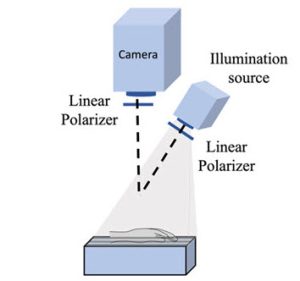
Spatial frequency domain imaging (SFDI) is the technique, where a light source obliquely illuminates the surface to be analysed and the camera take a perpendicular view.
In this case, the surface was the back of a person’s hand and both 880nm illumination and viewing were through linear-polarisers.
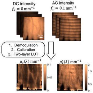
 Data is extracted as the change in return from place to place on the hand, expressed as a ‘frequency’ with respect to distance on the surface – so, not cycles/s like Hz, but cycles/mm – allowing a spatial spectrum to be created for the surface.
– Medical researchers have borrowed from electricity, and have adopted the term ‘DC’ for returns that do not change with position, and ‘AC’ for returns that do vary with position.
The captured images are demodulated and calibrated, and the diffuse reflectance data is converted into absorption and reduced scattering coefficients using a look-up table.
Boston University, Harvard Medical School, and Brigham and Women’s have teamed up to see if SFDI would reveal blood lipid levels, using three specific spatial wavelengths to evaluate hemoglobin, water and lipid concentrations.
They also tracked blood pressure, heart rate and, from blood samples, triglyceride, cholesterol and glucose levels.
“The results indicated that the optical absorption changes at specific wavelengths accurately correspond to variations in lipid concentrations,” according to the International Society for Optics and Photonic (SPIE), which has published results form the research in its journal Biophotonics Discovery.
A machine learning model was trained on the SFDI data, and can predict triglyceride levels to within 40mg/dL, according to SPIE.
“The research suggests that SFDI could serve as a promising alternative, allowing for easier monitoring of how meals affect cardiovascular health,” said Boston University researcher Professor Darren Roblyer. “Overall, these findings highlight the intricate relationship between diet, body response, and cardiovascular risk, suggesting a need for further exploration of non-invasive assessment methods.”
Medically, changes in blood nutrient and lipid levels after consuming a high-fat meal are indicators of current and future cardiovascular health, according to SPIE.
This research also revealed that high-fat meals lead to an increase in tissue oxygen saturation, while low-fat meals caused a decrease, “suggesting that dietary fat can affect not just overall health but also immediate physiological responses”, said the Society. Peak changes occurred three hours after eating, coinciding with a triglyceride spike.
For more, read ‘Enhanced peripheral tissue oxygenation and hemoglobin concentration after a high-fat meal measured with spatial frequency domain imaging‘ in Biophotonics Discovery.

